

Benign Oral Lesion Surgery in Weston, FL
Safe, precise removal of non-cancerous oral growths for comfort and peace of mind.
Understanding Benign Oral Lesion Surgery
Benign oral lesion surgery in Weston, FL involves the safe removal of non-cancerous growths that develop in or around the mouth. These lesions—such as fibromas, mucoceles, papillomas, and epulis—are typically harmless but may cause discomfort, interfere with eating or speaking, or simply cause concern about their appearance or growth pattern.
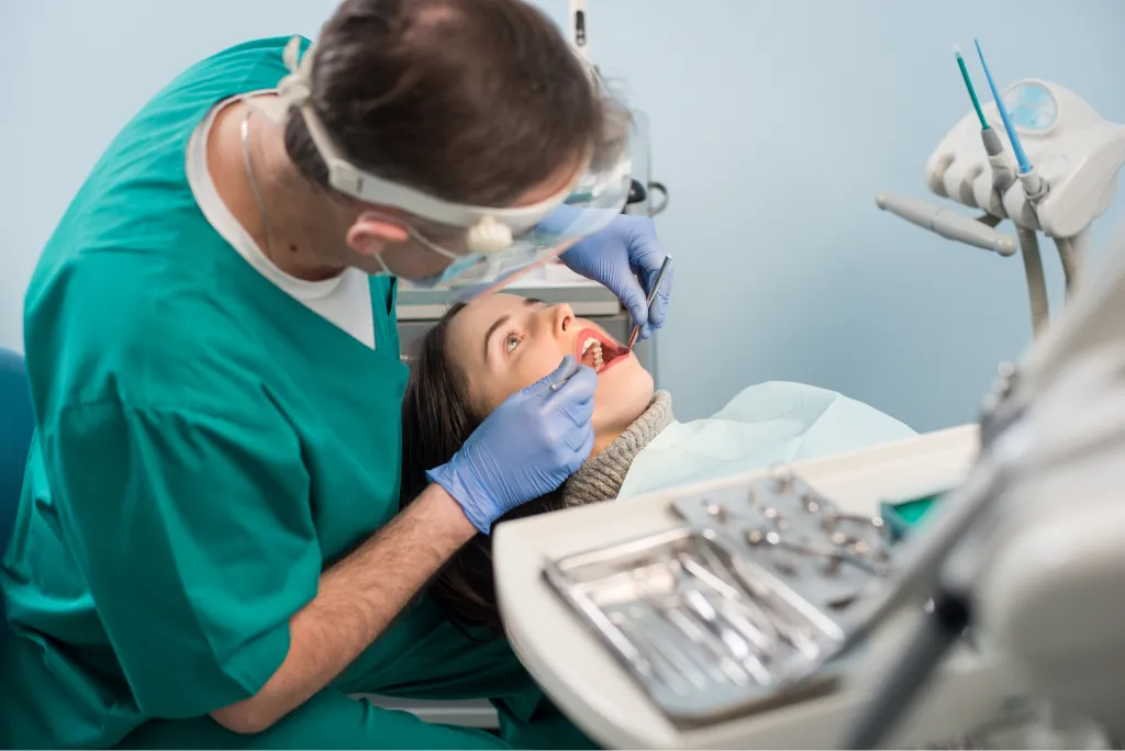
At Indian Trace Dental, Dr. Rodolfo Villa and Dr. Edgar Navarrete perform benign lesion excisions chairside under local anesthesia. Each case is approached with precision to remove the lesion completely while preserving surrounding healthy tissue. All excised tissue is sent for pathology analysis to confirm the diagnosis and provide peace of mind.
Common Types of Benign Oral Lesions
Fibromas are firm, painless lumps caused by chronic irritation. Mucoceles are fluid-filled cysts from blocked salivary glands. Papillomas are small, wart-like growths from viral exposure. Epulis are localized gum growths that may develop near ill-fitting dentures. Regardless of type, surgical removal provides definitive treatment and tissue for diagnostic confirmation. Patients from Weston, Davie, Pembroke Pines, and Sunrise trust Indian Trace Dental for careful oral lesion management.
Benefits of Benign Lesion Removal
A simple procedure that eliminates discomfort and confirms your oral health status.
Complete Removal
Surgical excision eliminates the lesion entirely with appropriate margins.
Pathology Confirmation
Removed tissue is analyzed by a lab to confirm benign status and rule out concerns.
Relieves Discomfort
Eliminates irritation from biting, chewing, or denture contact on the growth.
Quick Recovery
Most patients heal within one to two weeks with minimal post-operative care.
Low Recurrence
Proper excision technique results in a very low chance of the lesion returning.
In-Office Procedure
Performed chairside under local anesthesia with no hospital visit required.
The Lesion Removal Process
A straightforward in-office procedure with pathology confirmation.
Clinical Assessment
Your dentist examines the lesion, reviews your history, and may photograph or measure the area to plan excision.
Biopsy or Direct Excision
Depending on the lesion, an incisional biopsy may be performed first, or the entire lesion is excised in one procedure.
Local Anesthesia
The area is numbed completely to ensure a comfortable, pain-free surgical experience.
Surgical Removal
The lesion is carefully excised with a scalpel or laser, preserving surrounding healthy tissue. The site is closed with sutures if needed.
Pathology & Follow-Up
The specimen is sent for laboratory analysis. Results are reviewed with you within 7–14 days, and healing is monitored at a follow-up visit.
Related Diagnostic & Surgical Services
If a lesion needs evaluation before surgical removal, a soft tissue biopsy provides diagnostic clarity through laboratory analysis. We also perform frenectomy for restrictive tissue bands, wisdom teeth extraction, and implant placement as part of our comprehensive oral surgery services.
Regular dental checkups include oral screenings that help detect lesions early. If you notice any new growth, sore, or change in your mouth that does not resolve within two weeks, schedule an evaluation promptly.
Frequently Asked Questions About Benign Oral Lesion Surgery
Benign oral lesions are non-cancerous growths or abnormalities in the mouth. Common types include fibromas (firm tissue lumps), mucoceles (fluid-filled cysts), papillomas (wart-like growths), and epulis (gum growths). They are typically harmless but may cause discomfort or concern.
Removal may be recommended if the lesion interferes with eating, speaking, or denture fit; is frequently irritated or bitten; is growing in size; or needs to be examined by pathology to confirm it is not precancerous.
The procedure is performed under local anesthesia, so you will not feel pain during removal. Mild soreness at the site typically lasts a few days and is managed with over-the-counter pain medication.
Complete surgical excision has a very low recurrence rate for most benign lesions. Your dentist will remove the growth with appropriate margins and may recommend follow-up to monitor the area.
Yes. Excised tissue is routinely sent to a pathology laboratory for microscopic analysis to confirm the diagnosis and rule out any unexpected findings.
Most patients heal within one to two weeks. Soft diet, gentle rinsing, and avoiding irritation to the area help promote comfortable recovery.